Engineers on a Small Scale

Dentists and engineers have one thing in common: we build bridges!

My dad is a civil engineer, my brother is an architect. As expected, many people naturally speculated that I would follow their footsteps to pick up a career in engineering or construction. Here’s the plot twist: I didn’t really enjoy doing maths, but excelled in biology and chemistry. Ergo, the career option for engineering therefore went out the door. (I realise I theoretically could have done chemical engineering, but that’s up to an alternate universe to tell.)

IF YOU HAVE A MISSING TOOTH…

As a dentist I could still do one thing that an engineer does: design and build bridges! No complex calculations, just pure dentistry and an eye for aesthetics.

Bridges are one of three methods of replacing permanently lost teeth. The other two are dentures and implants, which I shall talk about on another day.

Here’s how dental bridges work…

 Dental bridge for tooth replacement.Image source

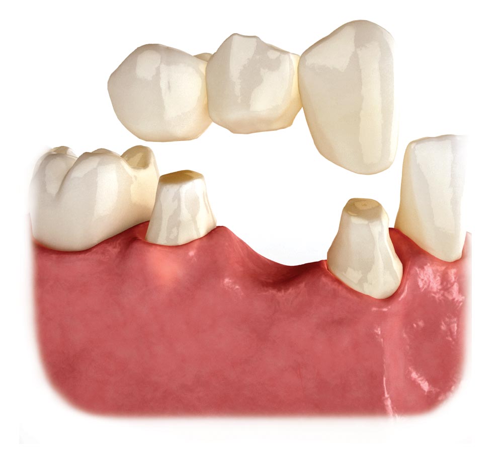
The picture above is an excellent illustration of a conventional dental bridge. There is a missing tooth down the middle. The two teeth beside it are drilled down in size and shaped to serve as abutments for the bridge to sit on.

An impression is taken of the drilled-down teeth and of the gums, and is sent to the dental lab to manufacture a bridge. If you know what dental crowns are and how they work, this is a similar thing: crowns joined together to form a bridge across the missing tooth gum region. The bridge is then cemented onto the abutment teeth and it would look as if the tooth was never missing in the first place.

NEW IS ALWAYS BETTER?

Image result for adhesive bridge

Image source

As you can tell from the previous example of a conventional bridge, the drill-down process of the natural teeth does a lot of unnecessary damage of the existing teeth. In modern day dentistry we try to conserve as much natural tooth as possible. So a new technique was used to “glue” bridges to the adjacent teeth – this is called adhesive bridges.

This requires only very minimal drilling to the back surfaces of the abutment teeth. After this, an impression is taken and sent to the lab, where they make the bridge as shown above – an artificial tooth (we call in a pontic) flanked by two metal wings. The wings are bonded to the adjacent teeth using a strong resin cement.

Adhesive bridges are much more commonplace these days than conventional bridges. They are less damaging to natural teeth and requires less chairside drilling. In fact, I just did one for my patient the other day, to replace a missing canine tooth! He went home a happy man, and can finally smile with confidence!

I thought it’d be interesting to share with everyone out there, explaining what dental bridges are and how they work. And also to talk a bit about my experience in giving a patient a bridge – it was a win-win!

Amalgam – A Story of Lustre and Glory

This is an article about amalgam – a story of lustre and  [REDACTED]  mercury.

Ah, the controversies and conflict of choice when it comes to picking a filling material. There have been numerous researches on which is a better filling material, and for what purposes. But I’m not going to delve into those: anyone can use Google and find out more. (But be warned! The internet is a hodgepodge of wacky and satirical information – read and learn with a pinch of salt!) I am, however, going to take on this topic from the patient’s point of view – and how, as a patient, you can make an informed decision.

So the other day I had a patient who had a mouthful of amalgam (silver fillings). He told me he wanted to get them all out, to be replaced with composites (white fillings). He claimed that this was not because he wanted to look good – the amalgam fillings were all in the molar teeth, way behind the smile line – but because he was reading articles about the controversies of amalgam and health. This was clearly an informed patient who knew quite a lot of stuff. However, I could not guarantee him that his fillings would be replaced with composite – different teeth may require different material choices. I hope that he was not too disappointed.

So what is AMALGAM?

 Pic source

Amalgam is an alloy of metals containing silver, tin, copper, zinc and mercury. Wait, what! Mercury? The toxic material?

Relax, mercury in amalgam is not going to kill you. Amalgam has been around for a really long time. Scientific evidence, accumulated over decades, supports the view that there is no clinical evidence of mercury poisoning in people who have amalgam fillings in their mouths. [1] This is because the main exposure to mercury from dental amalgam occurs during placement or removal of restoration in the tooth. [2]

So in any case, if there were to be mercury poisoning, it would be the dentists – who are exposed to mercury and its vapour for many many years. So far so good, no old dentists seem to suffer from any form of mercury poisoning. This is because the levels of mercury in amalgam are so minimal they are unlikely to do any kind of damage to the human body. Nevertheless, if you have the time, read the research paper in link no. 2 about it at the references section below.

Ughh, but it’s still so ugly. Why do dentists still use amalgam!

Well, it depends on which tooth the cavity is, and how the cavity is shaped. Of course, your front teeth will definitely not be the ones having amalgam – we almost always have a consensus that composite white fillings are better. Nobody wants a piece of metal sticking out of their smile in a selfie!

However, the front teeth is hardly the place for decay to occur, requiring fillings. By far the most common tooth decay is seen on the biting surfaces of the molar teeth. And this is where amalgam is at its best.

It is a metal – so it can withstand strong biting forces. Molar teeth have extremely strong forces acting on each other when biting and chewing. And perhaps for importantly, it survives for a very long time. Studies have shown that amalgam survives longer than composite (in the molar teeth) and is less likely to fail as a filling. [3]

If the decay on a molar tooth is small, composites will be used instead. Amalgam requires some degree of cohesion in the cavity for it to function properly.

FUN FACT!: Placing an amalgam filling requires excellent hand carving skills. We need to carve the filling to try and match the original tooth shape. Dentistry is as much an art as it is a science.

—–

I am still in the midst of my crown course, and I will hopefully be blogging about crowns really soon!

References

[1] http://www.poison.org/articles/2010-dec/do-fillings-cause-mercury-poisoning

[2] http://www.ncbi.nlm.nih.gov/pmc/articles/PMC3388771/

[3] http://www.ncbi.nlm.nih.gov/pubmed/17545266

Drill n’ Fill

This is an article about tooth decay and how dentists can fix it.

Tooth decay (we call them dental caries) is the most common disease affecting humans. [1] It is more common in the developed world than in the developing world, simply because of higher sugar intakes. According to the World Health Organisation, almost all adults have had a case of tooth decay at some point in their lives. [2]

Our tooth is made of several hard substances – such as enamel and dentine. Fun fact! Enamel is the hardest material in the body, even harder than bone. It can chew and grind moderately hard food, but gets destroyed by sugars.

Scumbag teeth

Image source

Tooth decay happens when tooth enamel gets demineralised (calcium crystals and salts are lost). Tooth decay needs 4 factors to happen:

  • Bacteria
  • Tooth surface
  • Sugars
  • Time

If we don’t brush our teeth, with time, bacteria feed upon the sugars left on our tooth surface. As they do so, they produce acid that destroys enamel – thus causing decay and a cavity.

Enamel decay is largely preventable, in that brushing teeth and keeping up with good oral hygiene can prevent it from happening. Early enamel decay is also reversible, in that if you find out about it early – seeing some brown staining or white spots on surfaces of teeth – you can do something to fix it yourself. If you see this happening, that means your brushing technique needs improvement, and with sufficient brushing with fluoride toothpaste these stains or spots will heal by themselves.

Image result for pit and fissure caries

However, when nothing is done to stop enamel decay, the decay spreads into dentine, and that’s where problems happen. In dentine decay, a blur brownish discolouration is seen through the translucent enamel. [3] At this point, you will feel pain and sensitivity, as the dentine can send signals to the nerve, telling your brain that there is an infection in your tooth.


Yikes. So what do we do about tooth decay!?

As the title of this post suggests, we do drilling and filling.

Drill: We use a diamond drill to cut through the enamel and access the decayed cavity. And then we use a round steel drill to take out the decayed substances, clearing out an area leaving a gaping hole where the decayed material once was.

Fill: Depending on the situation and tooth and patient’s choices, we choose from a whole variety of different filling materials. Some examples of such materials are amalgam (silver metal filling), composite (white/tooth-coloured filling) and glass ionomer (white temporary filling). Different filling materials have different techniques of placing them in the tooth and I think I will be able to blog about each of these materials at a later time. But all of them have this in common: they must be shaped and moulded back to how the tooth originally looked like, so that it would look nice and would not affect the bite.

Tooth decay and fillings is one the earliest clinical techniques we learned. It started in 2nd year and will carry on till we, well, retire. As tooth decay is the commonest disease in the world we won’t be seeing any of these go away so it is the most basic and handy skill to acquire in our dental journey.

I hope you enjoyed the post and learned a lot from it. Remember to brush your teeth and avoid sugars!


References

[1] Dental caries is a preventable infectious disease

[2] WHO Oral Health fact sheet

[3] What Constitutes Dental Caries? Histopathology of Carious Enamel and Dentin Related to the Action of Cariogenic Biofilms

Phantom Heads

This is a post on how we were taught the basic hands-on skills in the dental school.


“I lay there every morning, dressed in sky blue, mouth wide open, for students to work on my teeth. They can make mistakes on me, I’m fine with that. I’d never complain. My rubbery cheeks contain the water spray, and the suction at the bottom of my throat drains the water away. I’d never choke or gag. I am the ideal experimental patient.” — Mr Phantom Head

Before we’re allowed to see actual patients, we had to go through this clinical techniques course in 2nd year, where we work on phantom heads as shown above. Here in the clinical techniques lab (CTL) we were familiarised with the instruments and basic dental skills. The skills acquired here were mainly on drilling & filling in 2nd year and root canal treatment in the first half of 3rd year. Most of these skills translate to actual procedures done on patients in the conservation clinic in 3rd year.

The CTL is a simulated environment, much like flight and military simulators. Here we were trained in the art and science of dentistry with little to no cross-infection risk or patient complications. Here we were taught to sharpen and hone our skills to near perfection and had to pass several competency tests. Only upon passing these tests were we allowed to see and treat actual human patients. The CTL training serves as a platform as we graduated from preclinical students to clinical students.

This picture above shows the plastic teeth and the attached “jaw”. The “jaws” are magnetised so that they can be attached to the phantom heads. In essence we’re working on plastic teeth on a plastic head with rubbery cheeks. The head can be moved about and placed at exactly the right spot. Despite being the closest simulation there is to actual patient contact, there are some aspects of dentistry that cannot be simulated. A few of these things are: the tongue moving about, the cheeks and lips being more obstructing than the rubber ones, and patient issues such as pain and sensitivity.

Despite already in clinics and treating patients, we are still required to use the CTL as we learn more and more skills in the upcoming years. I must say that the CTL is so very essential in our preparation prior to clinical education and is the best way, as far as we know, to train dental students before patient contact. However, treating human patients is a whole new ball game and that’s why we have three full years of clinical training in the dental course.